Dermatomyositis(DM) | Dermatomyositis(DM) is a rare connective-tissue disease which is an idiopathic , autoimmune and chronic inflammatory mostly involved skin and muscles. It’s a systemic disorders that can also affect the joints, the oesophagus, the lungs, the kidney ,the heart ,etc. It’s more common in elderly people (over 40 yrs).In children most often appears between 5 and 15 years. Female to male ratio is 2:1. Black more affected.
Causes of Dermatomyositis:
- Idiopathic , but can be result from viral infections or cancer, that might cause autoimmune response.
- Inherited, and HLA subtypes (HLA-DE3,HLA-DR52,HLA-DR6).
- Environmental factors
- Certain drugs like statins.
- Certain Autoimmune diseases
Risk factors:
- Genetic
- UV rays exposure(sun)
- Certain drugs eg: statins
- Environmental factors
- Viral and parasitic infections
- Epidemiological factors
Pathophysiology:
Dermatomyositis occurs as a result of a humoral attack against the muscle capillaries and small arterioles.the disease starts when putative antibodies or other factors activate C3, forming C3b and C4b fragments that leads to formation of C3bNEO and membrane attack complex, which are deposited in the endomysial vasculature.B cells and CD4 (helper) cells are also present in abundance in the inflammatory reaction associated with blood vessels.
As disease progresses , the capillaries are destroyed and the muscles undergo micro infarction. The pathogenesis of the cutaneous component of dermatomyositis is poorly understood, but assumed as similar to muscle involvement. It’s also by the direct cytotoxic effect of CD8+ lymphocytes on muscles. Others cytokines as well as also TNF association. Abnormal T cell activity also plays role to cause dermatomyositis.
Classification :
- Idiopathic inflammatory myopathy along with polymyositis.
- Necrotizing autoimmune myositis
- Cancer –associated myositis
- Sporadic inclusion body myositis.
Sign and Symptoms:
- Symmetrical proximal muscles weakness distal muscles preserved.
- Muscles fatigue and weakness when climbing stairs, walking, rising from a sitting positions, combing their hair or reaching for items in cabinets that are above theirs shoulders.
- Muscles tenderness
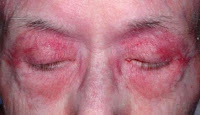
- Heliotrope rash ( blue-purple) discoloration around the eyes along with swelling, mainly upper eyelids .
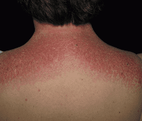
- Rash occurs on the upper chest or back i.e called “Shawl” (around neck) or “V- sign” above breasts.
- Rash present on face, upper thighs, upper arms, or hands.
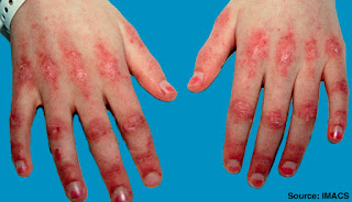
- Gottron’s sign (scaly eruptions rash on the knuckles )
- Periungual telangiectasia
- Around 30 percent of people have swollen ,painful but mild.
- Constitutionals symptoms like fever malaise ,weakness ,fatigue,etc.
- Nail changes (capillary loop dilations) , periungual erythema.
- “Mechanical Hands”-rough , crackels skin at tips and lateral aspects of the fingers resulting in irregular ,dirty appearing lines.
- Flagellated erythema around front trunk.

Heliotrope rash
 |
| shawl sign |
 |
| gottron's sign |
Others symptoms may include:
- Problem swallowing
- Lung problems
- Hard calcium deposits underneath the skin, that is mostly seen in children.
- Unintentional weight loss.
Note: ocular muscles never Involved.
Diagnostic Criteria:
Bohans and Peter criteria –
- Symmetric proximal muscles weakness .
- Typical rash (Heliotrope and gottron’s).
- Elevated serum muscle enzyme(CPK/aldolase).
- Myopathic changes on EMG.
- Characteristic muscle biopsy abnormalities and absence of histopathologic signs of other myopathies.
- Specific test for arthritis and arthralgia.
Diagnosis:
Lab: CBC: Increase inflammatory markers, anemia
Biochemical test: CPK /aldolase (best first step in diagnosis DM) are high, C-reactive increase.
Other tests: High result of AST (Asparted Aminotransferase) or Lactic Dehydrogenase(LDH), glutamate oxaloacetate ,pyruvate transaminase.Thyroid function test, electrolyte abnormality test( potassium,calcium,etc.), test for vitamin D
Clinical Urine Analysis(UA): abnormal in case of kidney affections.
Serology: + Antinuclear Antibody (ANA), Anti-Mi-2 antibodies (highly specific),Anti-Jo-1 (antihistidyl transfer RNA [tRNA].
Genetic testing: for HLA subtypes (HLA-DR3,HLA-DR52,HLA-DR6).
Muscle biopsy( gold standard and most accurate taste):
Under microscope and finding mononuclear white blood cells ,abnormal muscles cell degeneration and regeneration, dying muscles cells .
MRI (Magnetic resonance imaging): Guide muscle biopsy to investigate involvement of internal organs.
X-ray: Investigate joint involvement and calcifications.
Differential diagnosis:
- Polymyositis
- Polymyalgia rheumatic
- Electrolyte abnormalities ( potassium, calcium)
- Myasthenia gravis
- Eaton lambert syndrome
- Muscular dystrophy
- Infectious myositis
- Drug induced myositis
- Inclusion boby myositis
Treatment:
- Prednisone
- Azathioprine
- MTX
- Cyclophosphamide
- Cyclosporine
- Intravenous Immunoglobulin(IV Ig)
For skin treatment:
- Avoidance of sun exposure and sun protections measures including broad-spectrum sunscreens.
- Hydroxychloroquine and Chloroquine
For Calcinosis:
- Calcium channel blockers i.e diltiazam(240 mg bid) that’s help gradual resolution of calcinosis.
Note : female patients with dermatomyositis should be screened for ovarian cancer.
Complications of Dermatomyositis:
- Interstitial lung diseases
- Esophageal diseases
- Myocarditis, conductions disorders
- Trophic changes on skin (bed sores)
- Aspirated pneumonia
- Kidney abnormalities eg: Glomerulonephritis
- GIT abnormalities: dyspepsia, constipation, etc
- Respiration problem due to diaphragmatic muscle involvement
- Malignancies
Malignancy in Dermatomyositis Patients:
- 48 % >65 yr.
- 9% under 65 yr.
Types of cancer: adenocarcinoma of cervix, lung, ovaries, pancreases, bladder and stomach approx. 70% of associated cancers.
Cancer screening in Dermatomyositis Patients:
- Medical History and physical exam
- Age appropriate cancer screening(mammogram and colonoscopy)
- CT scan of chest, abdomen and pelvis in case of
- increased significant risk
- Pelvic USG and transvaginal USG for female
- Serum CA125 & CA 19-9
- PAS
- UA for blood
Prognosis of Dermatomyositis :
- Spontaneous remission in 20%
- 5% have fulminant progression and eventual death
- Poor prognostic factor –recalcitrant disease, delay in diagnosis, older age, malignancy, fever, asthenia-anorexia, pulmonary interstitial fibrosis
- Causes of death- malignancy, cardiac and pulmonary dysfunction and infection.
