A brain tumor or intracranial neoplasm occurs when abnormal cells form within the brain
Classification
Tumors of the Glial Tissue (Gliomas)
- Astrocytoma
- Oligodendroglioma
- Dendymoma
Tumors of the Neurons
- Gangliocytoma
- Neuroblastoma
- glioneurocytoma
- Giomatosis cerebri
- Cerebral neuroblastoma
- Central neurocytoma
Mixed tumors with Glial and Neuronal components
- Ganglioglioma
- Dysembryoplastic Neuroepithelial tumour (DNT)
Poorly differentiated and Embryonal Tumors
- Medulloblastoma
- Atypical teratoid/rhabdoid tumour
- Medulloepithelioma
- Dyspiastic Gangliocytoma of the cerebellum (Lhermitte-Duclos disease)
- Polar spongioblastoma
Tumors of the Meanings
- Meningioma
Peripheral nerve sheath tumors
- Schwannoma
- Neurofibroma
- Malignant nerve sheath tumors - MPNST(malignant schwannoma)
Miscellaneous Tumors
- Hemangioblastoma
- Craniopharyngioma
- Pituitary tumors Mesenchymal tumors
Metastatic tumors
- Primary CNS lymphoma
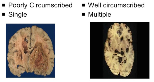
Difference between Primary &Secondary Brain Tumors
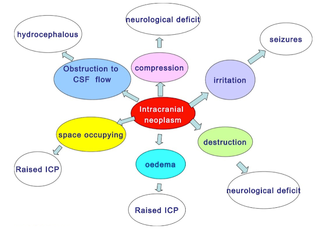
Pathophysiological affects of intracranial neoplasms
Fibrillary Astrocytic Neoplasms
Most Frequent
Infiltrative growth
Grade I - IV
Increased cellularity
Nuclear Pleomorphism
Mitotic activity
Endothelial proliferation
Necrosis - palisading
Clinical features
- Raised intracranial pressure
- Headache, nausea, vomiting
- Focal neurological signs
- weakness
- Seizures
Oligodendroglioma (Grade II/IV)
- Constitute 5% to 15% of gliomas .Fourth and fifth decade Cerebral hemispheres, with a predilection for white matter lorphology
- Grossly they are well-circumscribed, gelatinous, gray masses, often with cysts, focal hemorrhage, and calcification
- Microscopically the tumors are composed of sheets of regular cells with spherical nuclei containing finely granular chromatin surrounded by a clear halo of cytoplasm (fried egg appearance)
- The tumor typically contains a delicate network of anatomizing capillaries(chickenwire)
- Calcification seen in 90% of cases
Ependymomas (Grade II/IV)
- Arise next to the ependyma -lined ventricular system
- First two decades of life - they typically occur near the fourth ventricle
- In adults, the spinal cord is their most common location; jmors in this site are frequent in the setting of neurofibromatosis type 2
Gross
- Solid/papillary masses extending from the floor of the ventricle
- Variant Myxopapillary ependymomas occurs in the filum terminaie of the spinal cord.
NEURONAL TUMORS
- Central neurocytoma: low-grade neoplasm found within and adjacent to the ventricular system characterized by evenly spaced, round, uniform nuclei and often islands of neuropil
- Gangliogliomas are tumors with a mixture of glial elements, usually a low-grade astrocytoma, and mature-appearing neurons
- Dysembryoplastic neuroepithelial tumor is a distinctive, low-grade childhood tumor
Embryonal (Primitive) Neoplasms
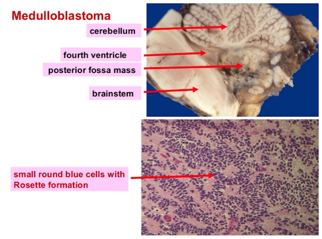
Medulloblastoma
- neuroectodermal origin, retain cellular features of primitive, undifferentiated cells.
- accounts for 20% of the brain tumors in children -Location exclusively in the cerebellum.
Morphology:-
- Grossly : well circumscribed, gray, and friable
- Microscopically : highly cellular and are composed of diffuse masses of small, undifferentiated oval or round cells, like a lymphoma
Rosette formation- are groups of tumor cells arranged in a circle around a fibrillary center
MENINGIOMAS (mostly Grade l/IV)
• predominantly benign tumors of adults, usually attached to the dura
• That arise from the meningothelial cell of the arachnoid.
Common sites of involvement
• Parasagittal aspect of the brain convexity
• Dura over the lateral convexity
• Wing of the sphenoid
• Olfactory groove, sella turcica
• Foramen magnum
• Ectopic meningioma
Macro:
- usually rounded masses, bosselated or polypoid appearance
- appearance
- Extension into the overlying bone may be present
- The surface of the mass is usually encapsulated
- Usually, they displace brain tissue without invading it
- Some meningiomas grow flat on the surface of the brain
- en plaque variant
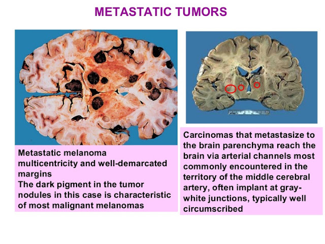
METASTATIC TUMORS
- Metastatic lesions, mostly carcinomas, account for approximately a quarter to half of intracranial tumors
- The five most common primary sites are lung, breast, skin (melanoma), kidney, and gastrointestinal tract, accounting for about 80% of all metastases.
- The Meninges are also a frequent site of involvement by metastatic disease.
- Intraparenchymal metastases form sharply demarcated masses often at the gray matter-white matter junction
- The boundary between tumor and brain parenchyma is well defined microscopically as well; melanoma is one tumor that does not always follow this rule
Schwannoma
• These benign tumors arise from the neural crest-derived Schwann cell and are associated with neurofibromatosis type 2.
• common location - cerebellopontine angle usually attached to vestibular branch of the eighth nerve
• Elsewhere within the dura, sensory nerves are preferentially involved, including branches of the trigeminal nerve and dorsal roots
- When extradural, most commonly found in association with large nerve trunks, where motor and sensory modalities are intermixed
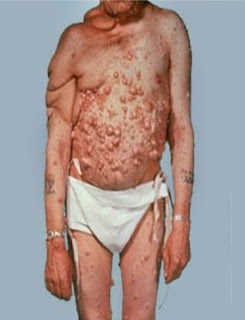
Neurofibromatosis
- an inherited disorder
- Affected individual develop multiple benign neurofibromas that arise within or are attached to the nerve trunks in the skin On the right side of the neck and shoulder of this patient, extensive subcutaneous neurofibromas have formed pendulous masses called plexiform neurofibromas
- an increased risk of developing neurofibrosarcomas
- This condition arises from mutations in the NF1 tumor suppressor gene
Treatment
- Treatment for brain tumors usually involves a combination of surgery, radiation and chemotherapy. The first goal of treatment is to remove as much of the tumor as possible, without damaging the surrounding normal brain tissue. Factors such as the patient's age, general health, occupation, and personal choice, all play a role in determining a course of treatment.
- Some nervous system tumors can be treated surgically and others cannot. Sometimes there are several different surgical procedures for a particular type of tumor. Sometimes a tumor can be treated with radiation alone, and does not need to be surgically removed.


Post a Comment
If you have any doubts, please let me know